Citriculturist
Aspiring
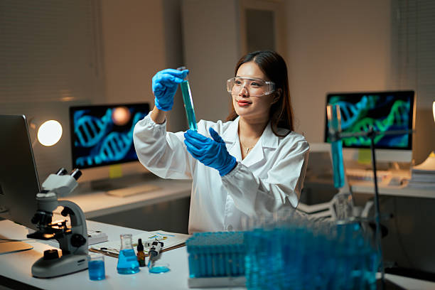
Explore CareerA Histologist is a specialized scientist or technician who studies the microscopic structure of tissues to understand biological processes, diagnose diseases, and support medical research. They work in hospitals, diagnostic laboratories, research institutions, universities, and forensic centers. Histologists apply staining techniques, microscopy, and molecular methods to prepare and analyze tissue samples, contributing to fields like pathology, medical diagnostics, and biomedical research. Combining expertise in biology, anatomy, and laboratory techniques, they play a crucial role in advancing healthcare, supporting clinical decisions, and contributing to scientific discoveries in a world where understanding tissue-level changes is vital for addressing health challenges.

A Histologist is a specialized scientist or technician who studies the microscopic structure of tissues to understand biological processes, diagnose diseases, and support medical research. They work in hospitals, diagnostic laboratories, research institutions, universities, and forensic centers. Histologists apply staining techniques, microscopy, and molecular methods to prepare and analyze tissue samples, contributing to fields like pathology, medical diagnostics, and biomedical research. Combining expertise in biology, anatomy, and laboratory techniques, they play a crucial role in advancing healthcare, supporting clinical decisions, and contributing to scientific discoveries in a world where understanding tissue-level changes is vital for addressing health challenges.
Histologists are experts who focus on the preparation, examination, and interpretation of tissue samples to identify cellular abnormalities, disease markers, and structural characteristics. Their work involves processing biological specimens, applying histological stains, using microscopes for detailed analysis, and documenting findings for medical or research purposes. They often operate in clinical, academic, or forensic settings, balancing technical precision with practical applications and interdisciplinary collaboration. Histologists are essential to fields like pathology, cancer research, and regenerative medicine, serving as experts in tissue science, contributing to solutions for accurate diagnoses, advancing medical research, and protecting scientific integrity, addressing pressing health issues through meticulous analysis, and improving outcomes through evidence-based strategies in a scientific landscape where histology’s relevance continues to grow due to advancements in diagnostics and personalized medicine.
Roles and Responsibilities:
Study Route & Eligibility Criteria:
| Route | Steps |
| Route 1 | 1. 10+2 with Physics, Chemistry, and Biology (PCB). 2. Bachelor’s degree in Biology, Biotechnology, or Medical Laboratory Technology (3-4 years). 3. Master’s degree in Histology, Pathology, or Biomedical Science (2 years). 4. PhD in Histology or related field (3-5 years, optional). 5. Postdoctoral research or clinical experience in histology labs (optional). |
| Route 2 | 1. 10+2 with Physics, Chemistry, and Biology (PCB). 2. Bachelor’s degree in Life Sciences or Zoology (3-4 years). 3. Master’s degree in Histology or Biomedical Science (2 years). 4. Specialized training in histological techniques (6 months-1 year). 5. Practical experience in diagnostic or research labs. |
| Route 3 | 1. 10+2 with Physics, Chemistry, and Biology (PCB). 2. Bachelor’s degree in Medical Laboratory Technology or Applied Sciences (3-4 years). 3. Master’s degree in Histology or Pathology (2 years). 4. Internship or fellowship in histology labs (1-2 years). 5. Certification in advanced histological techniques (optional). |
| Route 4 | 1. 10+2 with Physics, Chemistry, and Biology (PCB). 2. Bachelor’s degree from India in relevant field (3-4 years). 3. Master’s or PhD in Histology abroad (2-5 years). 4. Training or postdoctoral research in international histology programs (1-3 years). 5. Certification or licensure for international practice (if applicable). |
Significant Observations (Academic Related Points):
Internships & Practical Exposure:
Courses & Specializations to Enter the Field:
Top Institutes for Histologist Education (India):
| Institute | Course/Program | Official Link |
| All India Institute of Medical Sciences (AIIMS), New Delhi | MSc/PhD in Pathology | https://www.aiims.edu/ |
| Jawaharlal Nehru University (JNU), New Delhi | MSc/PhD in Life Sciences | https://www.jnu.ac.in/ |
| University of Delhi, Delhi | MSc/PhD in Biomedical Science | https://www.du.ac.in/ |
| Banaras Hindu University (BHU), Varanasi | MSc/PhD in Zoology | https://www.bhu.ac.in/ |
| Indian Institute of Technology (IIT), Kharagpur | MSc/PhD in Biotechnology | https://www.iitkgp.ac.in/ |
| University of Calcutta, Kolkata | MSc/PhD in Physiology | https://www.caluniv.ac.in/ |
| Savitribai Phule Pune University, Pune | MSc/PhD in Biotechnology | https://www.unipune.ac.in/ |
| Anna University, Chennai | MSc/PhD in Medical Biotechnology | https://www.annauniv.edu/ |
| University of Hyderabad, Hyderabad | MSc/PhD in Animal Biology | https://www.uohyd.ac.in/ |
| Christian Medical College (CMC), Vellore | MSc in Medical Laboratory Technology | https://www.cmch-vellore.edu/ |
Top International Institutes:
| Institution | Course | Country | Official Link |
| Harvard University | PhD in Pathology | USA | https://www.harvard.edu/ |
| Johns Hopkins University | MSc/PhD in Pathology | USA | https://www.jhu.edu/ |
| University of Oxford | DPhil in Pathology | UK | https://www.ox.ac.uk/ |
| University of Toronto | MSc/PhD in Laboratory Medicine and Pathobiology | Canada | https://www.utoronto.ca/ |
| University of Melbourne | MSc/PhD in Pathology | Australia | https://www.unimelb.edu.au/ |
| University of Cambridge | PhD in Pathology | UK | https://www.cam.ac.uk/ |
| Stanford University | PhD in Pathology | USA | https://www.stanford.edu/ |
| University of British Columbia (UBC) | MSc/PhD in Pathology and Laboratory Medicine | Canada | https://www.ubc.ca/ |
| Karolinska Institutet | MSc/PhD in Pathology | Sweden | https://www.ki.se/ |
| University of California, San Francisco (UCSF) | PhD in Biomedical Sciences | USA | https://www.ucsf.edu/ |
Entrance Tests Required:
India:
International:
Ideal Progressing Career Path
Undergraduate Student → Graduate Trainee (Master’s) → Junior Histologist → Established Histologist → Senior Histologist/Research Lead → Program Director/Professor
Major Areas of Employment:
Prominent Employers:
| India | International |
| All India Institute of Medical Sciences (AIIMS) | Mayo Clinic, USA |
| Apollo Hospitals | Johns Hopkins Hospital, USA |
| Fortis Healthcare | Cleveland Clinic, USA |
| Christian Medical College (CMC), Vellore | Massachusetts General Hospital, USA |
| Tata Memorial Hospital | Royal College of Pathologists, UK |
| Post Graduate Institute of Medical Education and Research (PGIMER) | National Institutes of Health (NIH), USA |
| King George’s Medical University (KGMU) | World Health Organization (WHO) |
| National Institute of Pathology (ICMR) | Centers for Disease Control and Prevention (CDC), USA |
| Medanta - The Medicity | Stanford Health Care, USA |
| Manipal Hospitals | University College London Hospitals (UCLH), UK |
Pros and Cons of the Profession:
| Pros | Cons |
| Significant contribution to medical diagnostics and research through tissue analysis. | Requires continuous learning to keep up with evolving histological and molecular techniques. |
| Intellectually stimulating work combining biology, pathology, and laboratory science in histological analysis. | Competitive field for clinical and research positions, often requiring extensive training and certifications. |
| High impact on patient care by contributing to accurate disease diagnosis and treatment. | Repetitive nature of laboratory work can lead to fatigue or burnout over time. |
| Opportunities for innovation in digital pathology and molecular histology techniques. | Limited public awareness of histology as a career, leading to fewer mainstream opportunities in some regions. |
| Growing relevance due to increasing demand for precise diagnostics and personalized medicine. | Potential exposure to biohazards and chemicals in laboratory settings, posing health risks. |
Industry Trends and Future Outlook:
Salary Expectations:
| Career Level | India (₹ per annum) | International (US$ per annum) |
| Trainee/Graduate Student | 2,50,000 - 4,50,000 | $25,000 - $35,000 |
| Junior Histologist | 4,50,000 - 8,00,000 | $40,000 - $55,000 |
| Established Histologist | 8,00,000 - 15,00,000 | $55,000 - $75,000 |
| Senior Histologist/Research Lead | 15,00,000 - 25,00,000 | $75,000 - $100,000 |
| Program Director/Professor | 25,00,000 - 40,00,000 | $100,000 - $140,000 |
Key Software Tools:
Professional Organizations and Networks:
Notable Histologists and Industry Leaders (Top 10):
Advice for Aspiring Histologists:
A career as a Histologist offers a unique opportunity to impact healthcare, medical research, and forensic science by studying the microscopic details of tissues. From preparing and analyzing tissue samples to supporting critical diagnoses and research breakthroughs, Histologists play a pivotal role in addressing some of the world’s most pressing health and scientific challenges. This field combines meticulous laboratory work, technological innovation, and interdisciplinary collaboration, offering diverse paths in clinical diagnostics, academia, and research. For those passionate about biology, tissue science, and medical discovery, a career as a Histologist provides a deeply rewarding journey with significant potential for making meaningful contributions to society in an era where histological analysis continues to shape diagnostic precision and therapeutic advancements.
Undergraduate students complete foundational education in biology or medical laboratory technology, learning basic sciences such as anatomy and...
0.0LPA
Trainees in Master’s programs focus on advanced studies in histology, learning tissue preparation and staining techniques under supervision. They...
0.0LPA
Early-career histologists establish roles in diagnostic labs, hospitals, or research facilities while developing their expertise and project...
0.0LPA
Take the next step and explore more about Histologist.
Explore similar career paths that might interest you